An MRI (magnetic resonance imaging) scan uses a strong magnetic field and radio waves to take pictures of the inside of your body. MRI scanning can be used to get detailed pictures of the heart, lungs and surrounding blood vessels and tissues.
MRI scanning can be used to detect blood clots in the lungs. It can also be used to show if the heart is working properly.
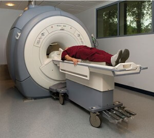
During an MRI scan, you lie on a bed that then slides into the scanner. The MRI scanner is like a tunnel about 1.5 metres long. You have to lie very still while each picture is being taken.
MRI scanners are very noisy, so you will be given headphones or earplugs to wear to protect your ears from the noise. You can listen to the radio through the headphones or bring a CD to listen to, if you want to.
If you are having an MRI scan you will be asked about any metal in your body which may interfere with the scan. This includes pacemakers, metal plates and pins, hearing aids and false eyes. It is important that you tell your healthcare team about any possible metal items inside your body before you have an MRI scan.
Watch a video of someone having an MRI scan, taken from the Pulmonary Hypertension Association (PHA UK) DVD Understanding Pulmonary Hypertension – a guide to diagnosis and treatment, here.